Comprehensive Eye Care
Eye Check-Ups and Tests
There are a number of tests which are performed to inspect the health vision of your eye. Our ophthalmologists decide the tests you need after examining the condition of your eyes.
Vision Screening
(FOR GLASSES, LENSES AND LASIK)
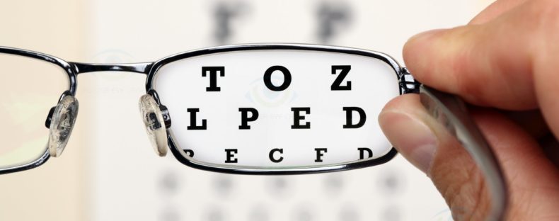
Visual Acuity Test
Refraction
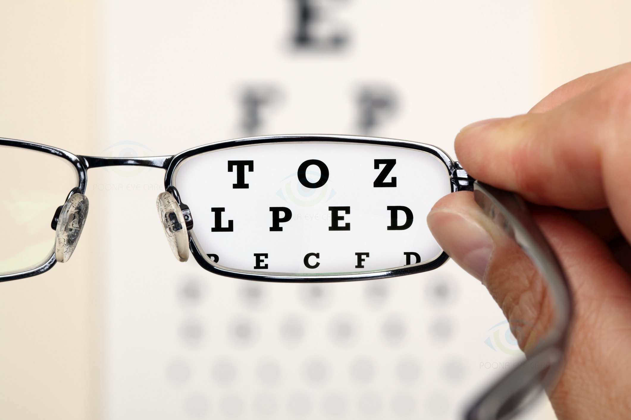
Visual Acuity Test measures the sharpness of your vision. It uses an eye chart to measure how well you can read a series of letters. Your eyes are tested one at a time, one is kept open to read, while the other eye is covered. Using a chart or a viewing device with progressively smaller letters, your eye doctor determines if you have 6/6 vision or if your vision shows signs of impairment.
The visual acuity test is a diagnostic indicator for many eye conditions involving sight from need for eye-glasses to severe conditions involving loss of vision.
If you have difficulty in reading, the doctor will then use a test called refraction. Refraction test uses several options of lenses in both eyes to determine the lens power you need. We also use an autorefractor or aberrometer to automatically estimate the eyeglass prescription. An aberrometer uses advanced wavefront technology to detect even obscure vision errors based on the way light travels through your eye.
General Check-Up
Color Blindness Test
Ocular Motility (Eye Movements) Testing
Stereopsis (Depth Perception) Test
Slit Lamp Examination

Color Blindness Test
This screening test checks your colour vision . A test called the Ishihara test is used to check colour blindness. It’s a simple test and is also available on the Internet.
Ocular Motility (Eye Movements) Testing
This test determines your eyes’ ability to follow moving objects. Your doctor will use a hand-held light and ask you to follow its movement. This test is important for children. Problems with eye movements can cause eye strain and affect the child’s performance in sports, reading and other areas.
Stereopsis (Depth Perception) Test
This test examines the 3D perception of your sight. It’s a simple test where circular patterns are presented to you. You have to identify the circles which are closest to you.
Slit Lamp Examination
A slit lamp is a binocular microscope which examines the structures of your eye under high magnification. It allows your eye doctor to see the structures at the front of your eye under magnification. The microscope is called a slit lamp because it uses an intense line of light, a slit, to illuminate your cornea, iris, lens, and the space between your iris and cornea. The slit allows your doctor to view these structures in small sections, which makes it easier to detect any tiny abnormalities.
A wide range of eye conditions and diseases can be detected with the slit lamp examination, including cataracts, macular degeneration, corneal ulcers and diabetic retinopathy etc.
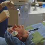
Dilated Eye Examination (Opthalmoscopy)
Doctors put drops to dilate the pupils. It takes about half an hour for the pupils to get substantially dilated. The doctors then use various instruments to check inside your eye. A dilated eye exam allows a doctor to examine your retina, optic nerve and other eye-structures with the help of an opthalmoscope.
A dilated eye exam is crucial to understand the health of your eye as many common eye-diseases have no early symptoms.
Diagnostic Tests
Glaucoma Tests
Cataract Tests
Tests For Lasik And Presbyond
Tests For Diabetic Retinopathy
Eye-Tests For Children

Along with your medical history, the doctor will ask for the following tests:
Tonometry
The first test for glaucoma is Tonometry, which is used to measure the pressure in your eyes. The doctor may sometimes use a yellow dye cum numbing agent for this purpose. The slit lamp and tonometer are used to perform tonometry. The test takes just a few seconds.
Optical Coherence Tomography
OCT in a non-invasive imaging test which examines the optic nerve.
Visual Field Test and Perimetry
This test is used to check blind spots inside the eye. These spots or stromas in the side-vision (peripheral vision) originate due to glaucoma. These spots can also help to understand areas of brain damage.Analysis of blind spots also may help identify specific areas of brain damage caused by a stroke or tumor. Perimetry is the systemic measurement of visual field function.
Gonioscopy
Gonioscopy is done to evaluate the internal drainage system of the eye, also referred to as the anterior chamber angle. The examination of this angle helps the doctor to determine the blockage in the flow of the aqueous fluid. Blockage leads to increase in eye-pressure which causes damage to the optic nerve, giving rise to glaucoma. Gonioscopy is done with the help of the split lamp.
Pachymetry
Pachymeter is a medical device used to measure the thickness of the cornea.
To determine whether you have a cataract, your doctor will review your medical history and symptoms, and perform an eye examination. Your doctor may conduct several tests, including:
Visual Acuity Test
Visual Acuity Test measures the sharpness of your vision. It uses an eye chart to measure how well you can read a series of letters. Your eyes are tested one at a time; one is kept open to read, while the other eye is covered. Using a chart or a viewing device with progressively smaller letters, your doctor will determine if you have 6/6 vision or if your vision shows signs of impairment.
Slit Lamp Test
A slit lamp is a binocular microscope which examines the structures of your eye under high magnification. It allows your eye doctor to see the structures at the front of your eye under magnification. The microscope is called a slit lamp because it uses an intense line of light, a slit, to illuminate your cornea, iris, lens, and the space between your iris and cornea. The slit allows your doctor to view these structures in small sections, which makes it easier to detect any tiny abnormalities. This helps doctor to detect clouding in the natural lens of your eye.
Retinal Examination Through Dilated Pupils with Slit Lamp
To prepare for a retinal exam, your eye doctor puts drops in your eyes to open your pupils wide (dilate). This makes it easier to examine the back of your eyes (retina). Using a slit lamp doctor will examine your lens for signs of a cataract.
IOL Master
IOL Master uses a scanning device to determine the required power for the intraocular lens to be implanted. It is a laser system which evaluates the length of the eye, surface curvature and anterior chamber depth for accuracy.
The success of Lasik surgery is directly related to how well you fit the criteria for being a good Lasik/Presbyond candidate.Tests are conducted to ascertain if you are the right candidate.
Visual Acuity Test
Visual Acuity Test measures the sharpness of your vision. It uses an eye chart to measure how well you can read a series of letters. Your eyes are tested one at a time, one is kept open to read, while the other eye is covered. Using a chart or a viewing device with progressively smaller letters, your eye doctor determines signs of vision impairment.
If the doctor finds it necessary, an examination called LVCA- Low Contrast vision Acuity Test and a test called FACT are also performed.
Refraction
Refraction test uses several options of lenses in both eyes to determine the lens power you need. We also use an autorefractor or aberrometer to automatically estimate the eyeglass prescription. An aberrometer uses advanced wavefront technology to detect even obscure vision errors based on the way light travels through your eye.
Keratometry (measurement of corneal curvature)
Keratometry (K) is the measurement of the corneal curvature; corneal curvature determines the power of the cornea. Differences in power across the cornea (opposite meridians) results in astigmatism; therefore, keratometry measures astigmatism.Astigmatism is one of the conditions which is corrected by Lasik treatment.
Slit Lamp Examination
A slit lamp is a binocular microscope which examines the structures of your eye under high magnification. It allows your eye doctor to see the structures at the front of your eye under magnification. The microscope is called a slit lamp because it uses an intense line of light, a slit, to illuminate your cornea, iris, lens, and the space between your iris and cornea. The slit allows your doctor to view these structures in small sections, which makes it easier to detect any tiny abnormalities.
Pupillary Measurements
The size of your pupils (pupillary measurements) is a very important eligibility criterea for Lasik and Presbyond surgeries. You cannot be eligible for lasik if the size of your pupils is large.
Tear Film Analysis
This test investigates the volume and quality of the tear film and health of the front eye. It documents the functions of eyelids, eyelashes, sclera, episclera, palpebral conjunctiva, tear ducts, tear glands etc. It uses slit lamp in conjunction with anterior eye imaging.
Pachymetry
Pachymeter is a medical device used to measure the thickness of the cornea.
Dilated Eye Examination (Opthalmoscopy)
Doctors put drops to dilate the pupils. It takes about half an hour for the pupils to get substantially dilated. The doctors then use various instruments to check inside your eye. A dilated eye exam allows your doctor to examine your retina.
Corneal Topography
Corneal topography is a computer assisted diagnostic tool that creates a three-dimensional map of the surface curvature of the cornea. The greatest advantage of corneal topography is its ability to detect irregular conditions invisible to most conventional testing. For laser vision correction, the corneal topography map is used in conjunction with other tests to determine exactly how much corneal tissue will be removed to correct vision and with what ablation pattern.
Penatcom Test
The Pentacam Comprehensive Eye Scanner is a rotating camera that photographs both the anterior (front) and posterior (back) surfaces and other areas of the cornea (the front part of the eye). The Pentacam Scanner’s main advantage is that it provides more precise measurements of the central cornea than any other ocular measurement instrument currently available.
Glaucoma Test
If the doctor finds it necessary he/she may undertake testing for glaucoma before surgery. Refer to Glaucoma Tests for details.
Comprehensive Dilated Eye Examination
Diabetic retinopathy is best diagnosed with a comprehensive dilated eye examination. For this, drops are put in your eyes to widen (dilate) your pupils to allow a better view inside the eyes.
The doctor looks for the following:
- Abnormal blood vessels
- Swelling, blood or fatty deposits in the retina
- Growth of new blood vessels and scar tissue
- Bleeding in the clear, jelly-like substance that fills the center of the eye (vitreous)
- Retinal detachment
- Abnormalities in your optic nerve
- Evidence of cataracts
Visual Acuity Test
Visual Acuity Test measures the sharpness of your vision. It uses an eye chart to measure how well you can read a series of letters. Your eyes are tested one at a time; one is kept open to read, while the other eye is covered. Using a chart or a viewing device with progressively smaller letters, your eye doctor determines signs of vision impairment.
Tonometry
Tonometer is used to measure the pressure in your eyes. The doctor may sometimes use a yellow dye cum numbing agent for this purpose. The slit lamp and tonometer are used to perform tonometry. The test takes just a few seconds. Doctor checks for signs of glaucoma with this test.
To assess whether your child’s eyes are developing normally, the doctor will conduct the following tests:
Pupil responses evaluation
Check if pupil opens and closes properly in the presence or absence of light.
Fixate and follow test
To check if eyes are able to fixate on and follow a light as it moves.
Preferential looking
Cards that are blank on one side with stripes on the other side are used to attract the gaze of an infant to the stripes.
LEA symbols
LEA symbols are used for young children. These are like regular eye tests but have special symbols like apple, house, square and circle etc. in place of letters.
Retinoscopy
A light is focused into the eye to observe the reflection from the back of the eye (retina). This test helps to determine the child's eyeglass prescription.
Random dot stereopsis
Special patterns of dots and 3-D glasses are used to measure how well the child's eyes work together as a team.
Stereopsis (Depth Perception) Test
This test examines the 3D perception of sight. Circular patterns are presented to the child. He/she has to identify the circles which are closest to him/her.
Physical checking
The doctor checks for lazy eye problem, alignment of eyes and focusing ability.
Color Blindness Test
This screening test checks the colour vision . A test called the Ishihara test is used to check colour blindness.
Slit Lamp Examination
A slit lamp is a binocular microscope which examines the structures of your child's eye under high magnification. It allows the doctor to see the structures at the front of your eye under magnification. The microscope is called a slit lamp because it uses an intense line of light, a slit, to illuminate the cornea, iris, lens, and the space between your iris and cornea. The slit allows your doctor to view these structures in small sections, which makes it easier to detect any tiny abnormalities.
Other treatments at Poona Eye Care
Please do write to us if you have any more queries on this. We will be glad to assist.
CONTACT US
BLOGS